Highlighter is on. Drag to highlight text. Click a highlight to remove it.
ALL / Nephrology
Vesicoureteric reflux
Vesicoureteric reflux
Vesicoureteric reflux (VUR) is the abnormal backflow of urine from the bladder into the ureter and kidney. It is a relatively common abnormality of the urinary tract in children and predisposes to urinary tract infection (UTI), being found in around 30% of children who present with a UTI. As around 35% of children develop renal scarring it is important to investigate for VUR in children following a UTIPathophysiology of VUR
- ureters are displaced laterally, entering the bladder in a more perpendicular fashion than at an angle
- therefore shortened intramural course of the ureter
- vesicoureteric junction cannot, therefore, function adequately
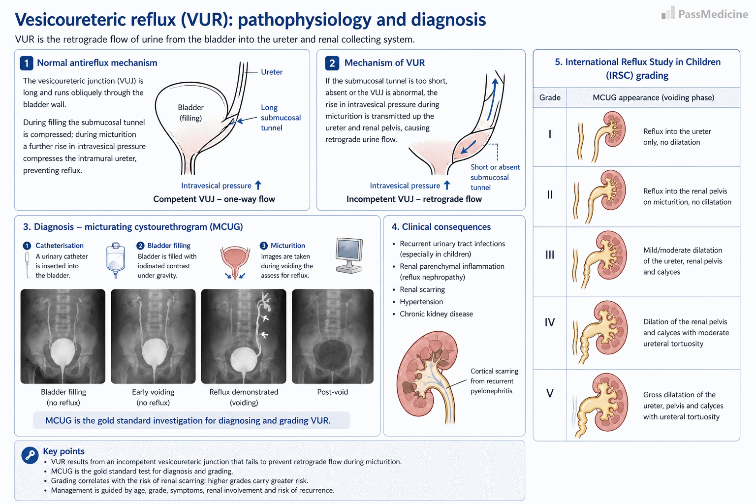
Pathophysiology and grading of vesicoureteric reflux
Possible presentations
- antenatal period: hydronephrosis on ultrasound
- recurrent childhood urinary tract infections
- reflux nephropathy
- term used to describe chronic pyelonephritis secondary to VUR
- commonest cause of chronic pyelonephritis
- renal scar may produce increased quantities of renin causing hypertension
Investigation
- VUR is normally diagnosed following a micturating cystourethrogram
- a DMSA scan may also be performed to look for renal scarring - this is typically located at the upper and lower pole and is associated with cortical thinning
The table below summarises the grading of VUR
| Grade | |
|---|---|
| I | Reflux into the ureter only, no dilatation |
| II | Reflux into the renal pelvis on micturition, no dilatation |
| III | Mild/moderate dilatation of the ureter, renal pelvis and calyces |
| IV | Dilation of the renal pelvis and calyces with moderate ureteral tortuosity |
| V | Gross dilatation of the ureter, pelvis and calyces with ureteral tortuosity |
+PassMedicine Notes
Vesicoureteric reflux (VUR) is the abnormal backflow of urine from the bladder into the ureter and kidney. It is a relatively common abnormality of the urinary tract in children and predisposes to urinary tract infection (UTI), being found in around 30% of children who present with a UTI. As around 35% of children develop renal scarring it is important to investigate for VUR in children following a UTI
Pathophysiology of VUR- ureters are displaced laterally, entering the bladder in a more perpendicular fashion than at an angle
- therefore shortened intramural course of the ureter
- vesicoureteric junction cannot, therefore, function adequately
- antenatal period: hydronephrosis on ultrasound
- recurrent childhood urinary tract infections
- reflux nephropathy
- term used to describe chronic pyelonephritis secondary to VUR
- commonest cause of chronic pyelonephritis
- renal scar may produce increased quantities of renin causing hypertension
- VUR is normally diagnosed following a micturating cystourethrogram
- a DMSA scan may also be performed to look for renal scarring - this is typically located at the upper and lower pole and is associated with cortical thinning
The table below summarises the grading of VUR
Grade_| I _ Reflux into the ureter only, no dilatation| II _ Reflux into the renal pelvis on micturition, no dilatation| III _ Mild/moderate dilatation of the ureter, renal pelvis and calyces| IV _ Dilation of the renal pelvis and calyces with moderate ureteral tortuosity| V_ Gross dilatation of the ureter, pelvis and calyces with ureteral tortuosity|