Ulcerative colitis
Ulcerative colitis
Ulcerative colitis (UC) is a form of inflammatory bowel disease characterised by relapsing inflammation of the colonic mucosa. It classically involves the rectum (hence it is the most common site for UC) and extends proximally in a continuous pattern. The extent varies from proctitis to pancolitis.Unlike Crohn's disease, UC does not usually cause skip lesions or involve the small bowel. However, limited terminal ileal inflammation may occur in extensive colitis ('backwash ileitis'). Rectal sparing or patchy inflammation may also occur, particularly following treatment and in children. The peak incidence of UC is in people aged 15-25 years and in those aged 55-65 years.
Clinical features
- symptoms usually develop gradually and follow a relapsing and remitting course
- bloody diarrhoea, often with mucus
- urgency
- tenesmus, particularly with proctitis
- frequent, small-volume stools
- cramping lower abdominal pain, commonly affecting the left lower quadrant
- systemic features in more severe disease: fever, tachycardia, weight loss and anaemia
- extra-intestinal manifestations (see below)
Initial investigations
- blood tests: FBC, CRP/ESR, U&E, LFTs and ferritin
- stool culture and testing for Clostridioides difficile to exclude infection
- faecal calprotectin may support the presence of intestinal inflammation but is not specific for UC
- diagnosis is confirmed using endoscopy with histological examination of biopsies
Endoscopy
- colonoscopy + biopsy is generally done for diagnosis
- however in patients with severe colitis colonoscopy should be avoided due to the risk of perforation - a flexible sigmoidoscopy is preferred
- typical findings:
- red, raw mucosa, bleeds easily
- no inflammation beyond submucosa (unless fulminant disease)
- widespread ulceration with preservation of adjacent mucosa which has the appearance of polyps ('pseudopolyps')
- inflammatory cell infiltrate in lamina propria
- neutrophils migrate through the walls of glands to form crypt abscesses
- depletion of goblet cells and mucin from gland epithelium
- granulomas are infrequent
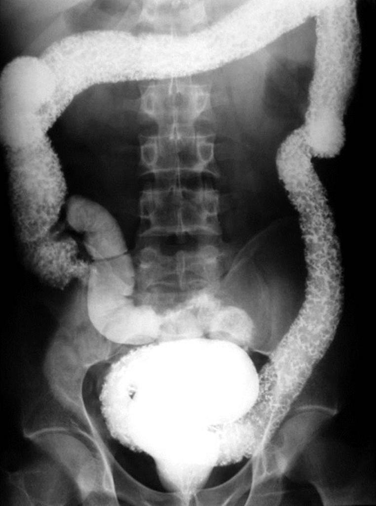
Barium enema
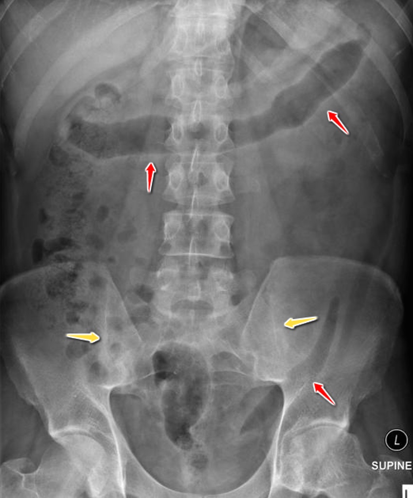
- loss of haustrations
- superficial ulceration, 'pseudopolyps'
- long standing disease: colon is narrow and short -'drainpipe colon'
Questions regarding the 'extra-intestinal' features of inflammatory bowel disease are common:
| Manifestations | Notes | |
|---|---|---|
| Usually parallels intestinal activity | Type 1 peripheral arthritis: acute, asymmetric and pauciarticular Erythema nodosum Episcleritis Oral aphthous ulceration | Peripheral arthritis is the most common extra-intestinal manifestation Type 1 arthritis usually affects large joints of the lower limbs |
| Usually independent of intestinal activity | Axial spondyloarthritis, including sacroiliitis and ankylosing spondylitis Type 2 peripheral arthritis: chronic, symmetrical and polyarticular Uveitis Primary sclerosing cholangitis | Primary sclerosing cholangitis is strongly associated with UC Uveitis may threaten vision and requires urgent ophthalmological assessment |
| Variable relationship to intestinal activity | Pyoderma gangrenosum | Pyoderma gangrenosum is associated particularly with UC, but its course does not reliably follow intestinal activity |
+PassMedicine Notes
Ulcerative colitis (UC) is a form of inflammatory bowel disease characterised by relapsing inflammation of the colonic mucosa. It classically involves the rectum (hence it is the most common site for UC) and extends proximally in a continuous pattern. The extent varies from proctitis to pancolitis.
Unlike Crohn's disease, UC does not usually cause skip lesions or involve the small bowel. However, limited terminal ileal inflammation may occur in extensive colitis ('backwash ileitis'). Rectal sparing or patchy inflammation may also occur, particularly following treatment and in children. The peak incidence of UC is in people aged 15-25 years and in those aged 55-65 years.
Clinical features- symptoms usually develop gradually and follow a relapsing and remitting course
- bloody diarrhoea, often with mucus
- urgency
- tenesmus, particularly with proctitis
- frequent, small-volume stools
- cramping lower abdominal pain, commonly affecting the left lower quadrant
- systemic features in more severe disease: fever, tachycardia, weight loss and anaemia
- extra-intestinal manifestations (see below)
- blood tests: FBC, CRP/ESR, U&E, LFTs and ferritin
- stool culture and testing for Clostridioides difficile to exclude infection
- faecal calprotectin may support the presence of intestinal inflammation but is not specific for UC
- diagnosis is confirmed using endoscopy with histological examination of biopsies
- colonoscopy + biopsy is generally done for diagnosis
- however in patients with severe colitis colonoscopy should be avoided due to the risk of perforation - a flexible sigmoidoscopy is preferred
- typical findings:
- red, raw mucosa, bleeds easily
- no inflammation beyond submucosa (unless fulminant disease)
- widespread ulceration with preservation of adjacent mucosa which has the appearance of polyps ('pseudopolyps')
- inflammatory cell infiltrate in lamina propria
- neutrophils migrate through the walls of glands to form crypt abscesses
- depletion of goblet cells and mucin from gland epithelium
- granulomas are infrequent
- loss of haustrations
- superficial ulceration, 'pseudopolyps'
- long standing disease: colon is narrow and short -'drainpipe colon'
Questions regarding the 'extra-intestinal' features of inflammatory bowel disease are common:
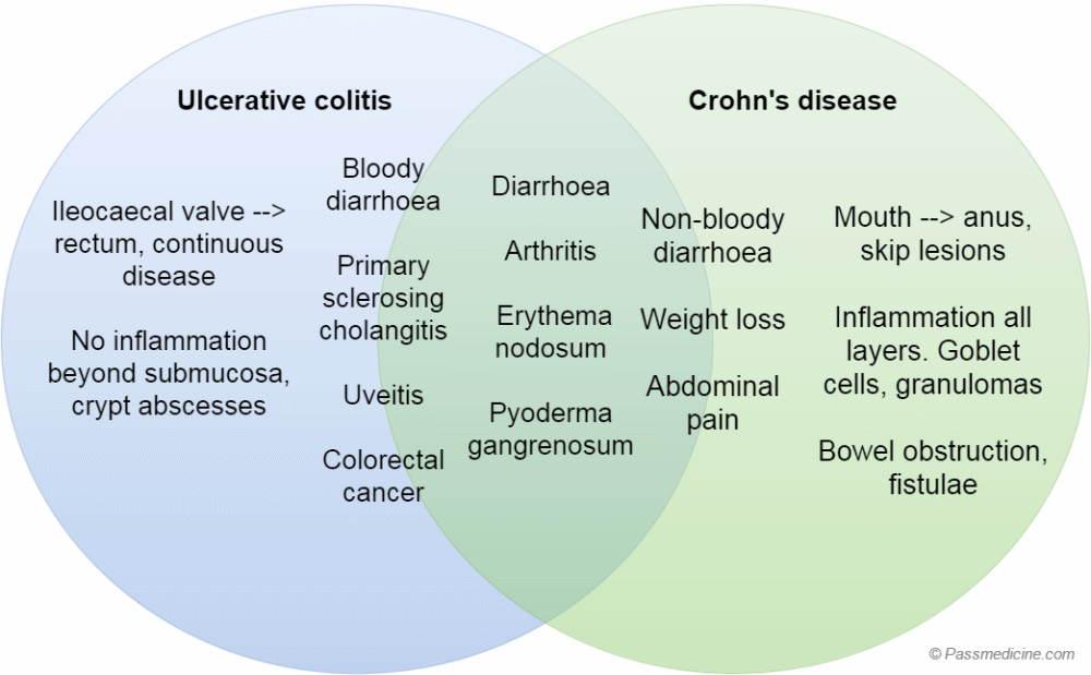
_ Manifestations _ Notes | Usually parallels intestinal activity _ Type 1 peripheral arthritis: acute, asymmetric and pauciarticular Erythema nodosum Episcleritis Oral aphthous ulceration _ Peripheral arthritis is the most common extra-intestinal manifestation Type 1 arthritis usually affects large joints of the lower limbs| Usually independent of intestinal activity _ Axial spondyloarthritis, including sacroiliitis and ankylosing spondylitis Type 2 peripheral arthritis: chronic, symmetrical and polyarticular Uveitis Primary sclerosing cholangitis _ Primary sclerosing cholangitis is strongly associated with UC Uveitis may threaten vision and requires urgent ophthalmological assessment| Variable relationship to intestinal activity _ Pyoderma gangrenosum _ Pyoderma gangrenosum is associated particularly with UC, but its course does not reliably follow intestinal activity| pdd910.png Venn diagram showing shared features and differences between ulcerative colitis and Crohn's disease. Note that whilst some features are present in both, some are much more common in one of the conditions, for example colorectal cancer in ulcerative colitis