Highlighter is on. Drag to highlight text. Click a highlight to remove it.
ALL / Neurology
Subarachnoid haemorrhage
Subarachnoid haemorrhage
A subarachnoid haemorrhage (SAH) is an intracranial haemorrhage that is defined as the presence of blood within the subarachnoid space, i.e. deep to the subarachnoid layer of the meninges.The most common cause of SAH is head injury and this is called traumatic SAH . In the absence of trauma, SAH is termed spontaneous SAH . The rest of this note focuses on spontaneous SAH.
Causes of spontaneous SAH include:
- intracranial aneurysm (saccular 'berry' aneurysms)
- accounts for around 85% of cases
- conditions associated with berry aneurysms include hypertension,adult polycystic kidney disease, Ehlers-Danlos syndrome and coarctation of the aorta
- arteriovenous malformation
- pituitary apoplexy
- mycotic (infective) aneurysms
Classical presenting features include:
- headache
- usually of sudden-onset ('thunderclap' or 'hit with a baseball bat')
- severe ('worst of my life')
- occipital
- typically peaking in intensity within 1 to 5 minutes
- there may be a history of a less-severe 'sentinel' headache in the weeks prior to presentation
- nausea and vomiting
- meningism (photophobia, neck stiffness)
- coma
- seizures
- ECG changes including ST elevation may be seen
- this may be secondary to either autonomic neural stimulation from the hypothalamus or elevated levels of circulating catecholamines
Investigation
- non-contrast CT head is the first-line investigation of choice
- acute blood (hyperdense/bright on CT) is typically distributed in the basal cisterns, sulci and in severe cases the ventricular system.
- if CT head is done within 6 hours of symptom onset and is normal
- new guidelines suggest not doing a lumbar puncture
- consider an alternative diagnosis
- if CT head is done more than 6 hours after symptom onset and is normal
- do a lumber puncture (LP)
- timing wise the LP should be performed at least 12 hours following the onset of symptoms to allow the development of xanthochromia (the result of red blood cell breakdown).
- xanthochromia helps to distinguish true SAH from a 'traumatic tap' (blood introduced by the LP procedure)
- xanthochromia is due to red blood cells in the CSF undergoing haemolysis: haemoglobin → oxyhaemoglobin → bilirubin. Spectrophotometry detects bilirubin and oxyhaemoglobin
- as well as xanthochromia, CSF findings consistent with subarachnoid haemorrhage include a normal or raised opening pressure
- if the CT shows evidence of a SAH
- referral to neurosurgery to be made as soon as SAH is confirmed
After spontaneous SAH is confirmed, the aim of investigation is to identify a causative pathology that needs urgent treatment:
- CT intracranial angiogram (to identify a vascular lesion e.g. aneurysm or AVM)
- +/- digital subtraction angiogram (catheter angiogram)
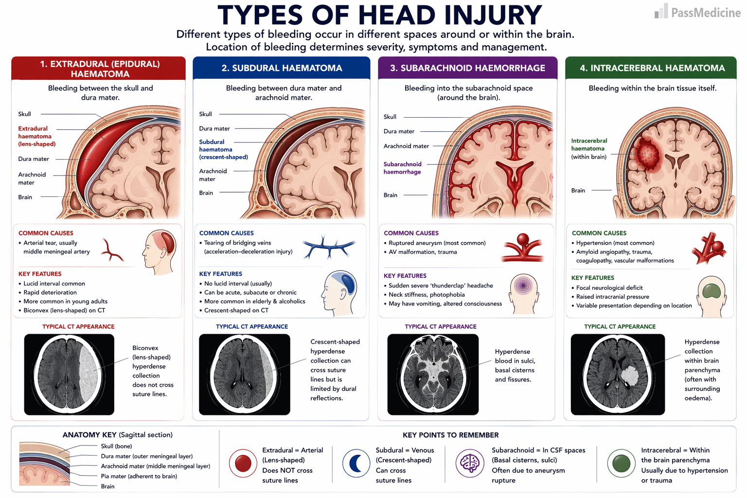
Types of head injury
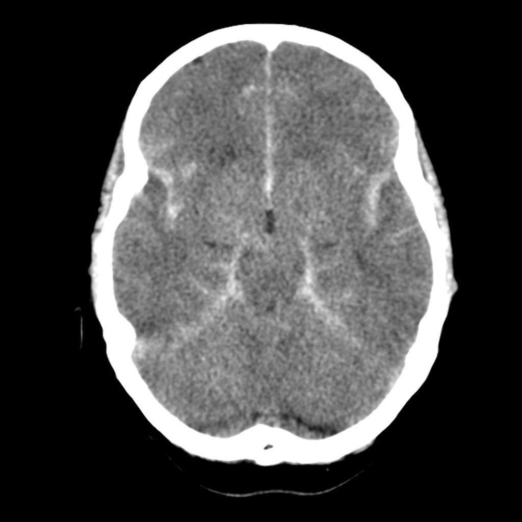
CT image shows diffuse subarachnoid haemorrhage in all basal cisterns, bilateral sylvian fissures and the inter-hemispheric fissure. This case demonstrates the typical distribution that takes the blood into the subarachnoid space in a subarachnoid hemorrhage.
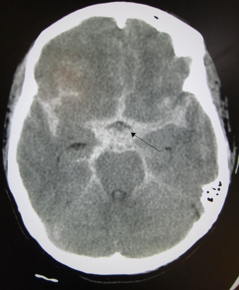
CT showing a subarachnoid haemorrhage as a white area in the centre (marked by the arrow) and stretching into the sulci to either side
Management of a confirmed aneurysmal subarachnoid haemorrhage
- supportive
- bed rest
- analgesia
- venous thromboembolism prophylaxis
- discontinuation of antithrombotics (reversal of anticoagulation if present)
- vasospasm is prevented using a course of oral nimodipine
- intracranial aneurysms are at risk of rebleeding and therefore require prompt intervention, preferably within 24 hours
- most intracranial aneurysms are now treated with a coil by interventional neuroradiologists, but a minority require a craniotomy and clipping by a neurosurgeon
Complications of aneurysmal SAH:
- re-bleeding
- happens in around 10% of cases and most common in the first 12 hours
- if rebleeding is suspected (e.g. sudden worsening of neurological symptoms) then a repeat CT should be arranged
- associated with a high mortality (up to 70%)
- hydrocephalus
- hydrocephalus is temporarily treated with an external ventricular drain (CSF diverted into a bag at the bedside) or, if required, a long-term ventriculoperitoneal shunt
- vasospasm (also termed delayed cerebral ischaemia), typically 7-14 days after onset
- ensure euvolaemia (normal blood volume)
- consider treatment with a vasopressor if symptoms persist
- hyponatraemia (most typically due to syndrome inappropriate anti-diuretic hormone (SIADH))
- seizures
Important predictive factors in SAH:
- conscious level on admission
- age
- amount of blood visible on CT head
+PassMedicine Notes
A subarachnoid haemorrhage (SAH) is an intracranial haemorrhage that is defined as the presence of blood within the subarachnoid space, i.e. deep to the subarachnoid layer of the meninges.
The most common cause of SAH is head injury and this is called traumatic SAH . In the absence of trauma, SAH is termed spontaneous SAH . The rest of this note focuses on spontaneous SAH.
Causes of spontaneous SAH include:- intracranial aneurysm (saccular 'berry' aneurysms)
- accounts for around 85% of cases
- conditions associated with berry aneurysms include hypertension,adult polycystic kidney disease, Ehlers-Danlos syndrome and coarctation of the aorta
- arteriovenous malformation
- pituitary apoplexy
- mycotic (infective) aneurysms
- headache
- usually of sudden-onset ('thunderclap' or 'hit with a baseball bat')
- severe ('worst of my life')
- occipital
- typically peaking in intensity within 1 to 5 minutes
- there may be a history of a less-severe 'sentinel' headache in the weeks prior to presentation
- nausea and vomiting
- meningism (photophobia, neck stiffness)
- coma
- seizures
- ECG changes including ST elevation may be seen
- this may be secondary to either autonomic neural stimulation from the hypothalamus or elevated levels of circulating catecholamines
- non-contrast CT head is the first-line investigation of choice
- acute blood (hyperdense/bright on CT) is typically distributed in the basal cisterns, sulci and in severe cases the ventricular system.
- if CT head is done within 6 hours of symptom onset and is normal
- new guidelines suggest not doing a lumbar puncture
- consider an alternative diagnosis
- if CT head is done more than 6 hours after symptom onset and is normal
- do a lumber puncture (LP)
- timing wise the LP should be performed at least 12 hours following the onset of symptoms to allow the development of xanthochromia (the result of red blood cell breakdown).
- xanthochromia helps to distinguish true SAH from a 'traumatic tap' (blood introduced by the LP procedure)
- xanthochromia is due to red blood cells in the CSF undergoing haemolysis: haemoglobin → oxyhaemoglobin → bilirubin. Spectrophotometry detects bilirubin and oxyhaemoglobin
- as well as xanthochromia, CSF findings consistent with subarachnoid haemorrhage include a normal or raised opening pressure
- if the CT shows evidence of a SAH
- referral to neurosurgery to be made as soon as SAH is confirmed
- CT intracranial angiogram (to identify a vascular lesion e.g. aneurysm or AVM)
- +/- digital subtraction angiogram (catheter angiogram)
- supportive
- bed rest
- analgesia
- venous thromboembolism prophylaxis
- discontinuation of antithrombotics (reversal of anticoagulation if present)
- vasospasm is prevented using a course of oral nimodipine
- intracranial aneurysms are at risk of rebleeding and therefore require prompt intervention, preferably within 24 hours
- most intracranial aneurysms are now treated with a coil by interventional neuroradiologists, but a minority require a craniotomy and clipping by a neurosurgeon
- re-bleeding
- happens in around 10% of cases and most common in the first 12 hours
- if rebleeding is suspected (e.g. sudden worsening of neurological symptoms) then a repeat CT should be arranged
- associated with a high mortality (up to 70%)
- hydrocephalus
- hydrocephalus is temporarily treated with an external ventricular drain (CSF diverted into a bag at the bedside) or, if required, a long-term ventriculoperitoneal shunt
- vasospasm (also termed delayed cerebral ischaemia), typically 7-14 days after onset
- ensure euvolaemia (normal blood volume)
- consider treatment with a vasopressor if symptoms persist
- hyponatraemia (most typically due to syndrome inappropriate anti-diuretic hormone (SIADH))
- seizures
- conscious level on admission
- age
- amount of blood visible on CT head