Renal anatomy
Renal anatomy
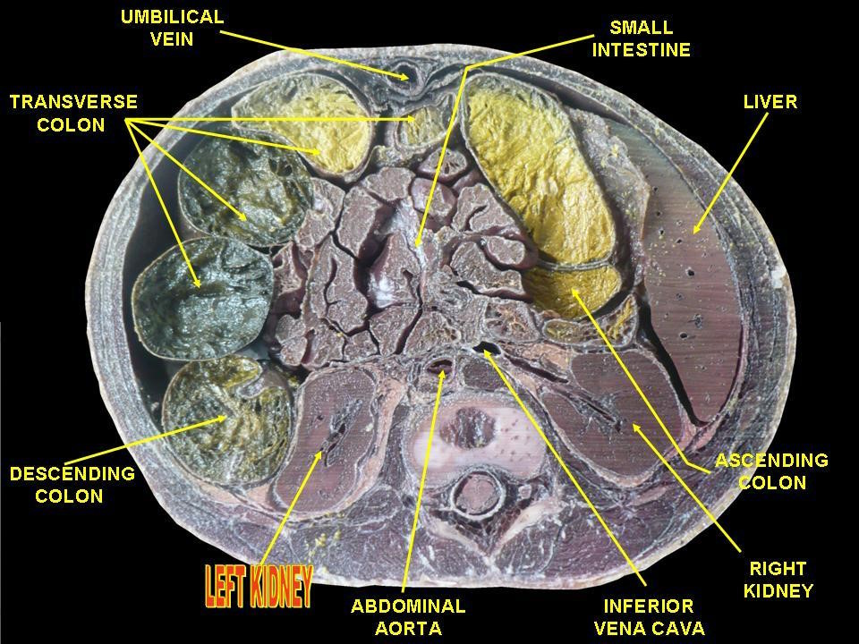
Each kidney is about 11cm long, 5cm wide and 3cm thick. They are located in a deep gutter alongside the projecting vertebral bodies, on the anterior surface of psoas major. In most cases, the left kidney lies approximately 1.5cm higher than the right. The upper pole of both kidneys approximates with the 11th rib (beware pneumothorax during nephrectomy). On the left hand side the hilum is located at the L1 vertebral level and the right kidney at level L1-2. The lower border of the kidneys is usually alongside L3.Relations
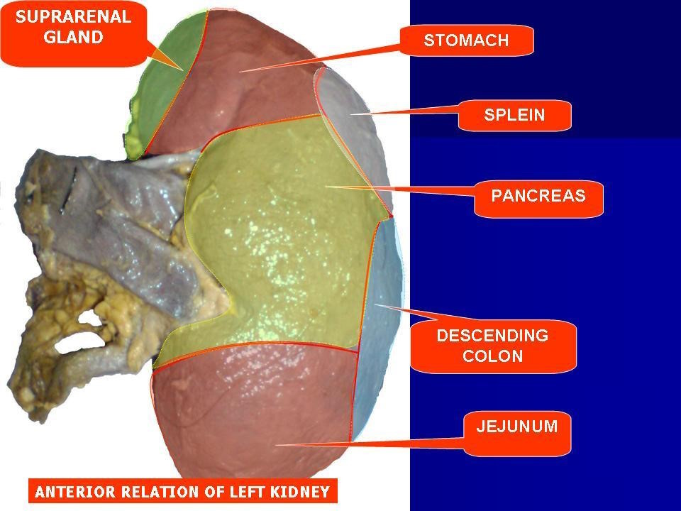
The tables below show the anatomical relations of the kidneys:
| Relations | Right Kidney | Left Kidney |
|---|---|---|
| Posterior | Quadratus lumborum, diaphragm, psoas major, transversus abdominis | Quadratus lumborum, diaphragm, psoas major, transversus abdominis |
| Anterior | Hepatic flexure of colon | Stomach, Pancreatic tail |
| Superior | Liver, adrenal gland | Spleen, adrenal gland |
If we consider relations according to whether they are in direct contact or whether there is peritoneum in-between:
Right kidney
| Direct contact | Layer of peritoneum in-between |
|---|---|
| Right suprarenal gland Duodenum Colon | Liver Distal part of small intestine |
Left kidney
| Direct contact | Layer of peritoneum in-between |
|---|---|
| Left suprarenal gland Pancreas Colon | Stomach Spleen Distal part of small intestine |
Fascial covering
Each kidney and suprarenal gland is enclosed within a common layer of investing fascia, derived from the transversalis fascia. It is divided into anterior and posterior layers (Gerotas fascia).
Renal structure
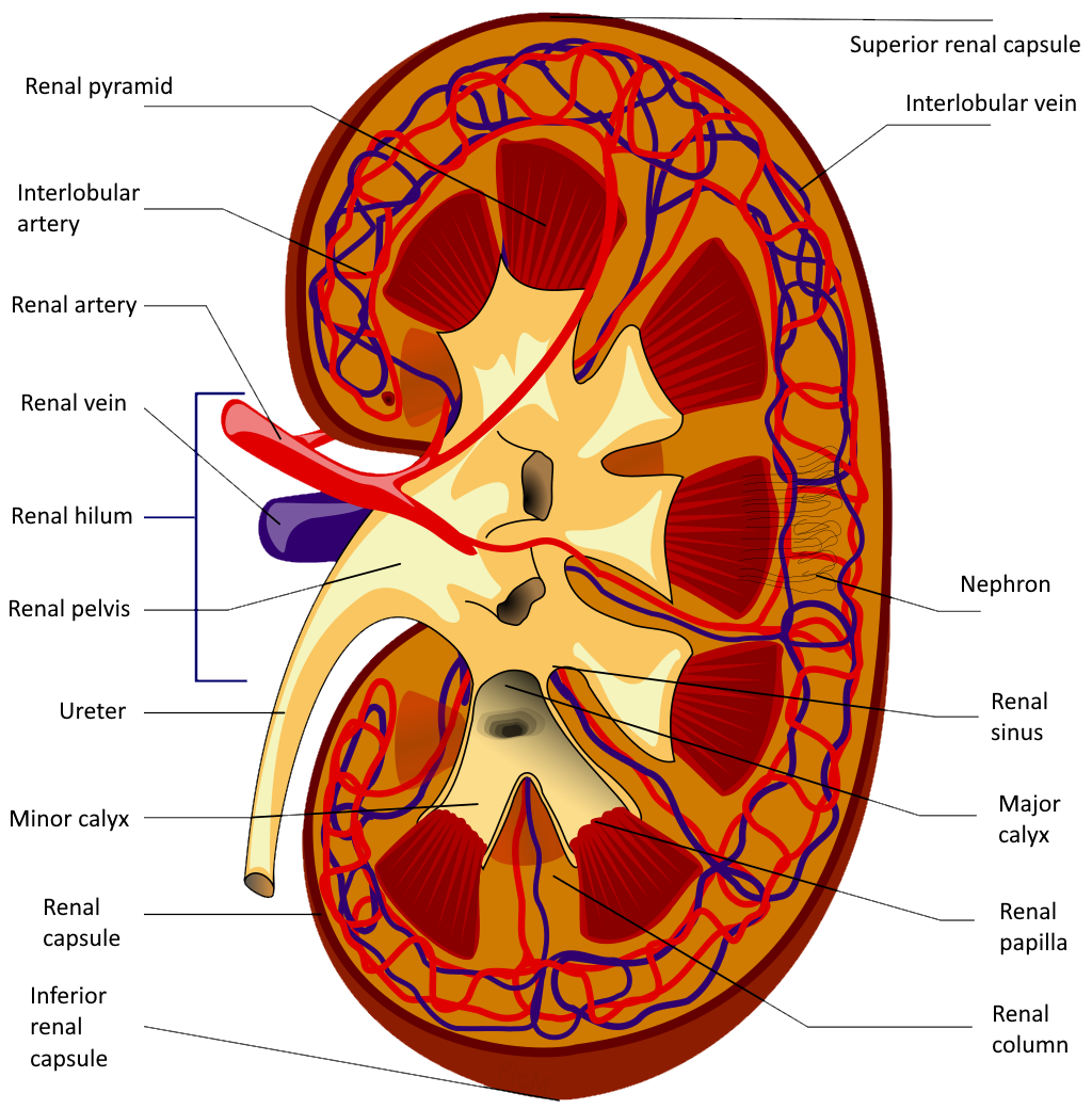
Kidneys are surrounded by an outer cortex and an inner medulla which usually contains between 6 and 10 pyramidal structures. The papilla marks the innermost apex of these. They terminate at the renal pelvis, into the ureter.
Lying in a hollow within the kidney is the renal sinus. This contains:
- 1. Branches of the renal artery
- 2. Tributaries of the renal vein
- 3. Major and minor calyces
- 4. Fat
Structures at the renal hilum
The renal vein lies most anteriorly, then the renal artery (it is an end artery) and the ureter lies most posteriorly.
+PassMedicine Notes
Each kidney is about 11cm long, 5cm wide and 3cm thick. They are located in a deep gutter alongside the projecting vertebral bodies, on the anterior surface of psoas major. In most cases, the left kidney lies approximately 1.5cm higher than the right. The upper pole of both kidneys approximates with the 11th rib (beware pneumothorax during nephrectomy). On the left hand side the hilum is located at the L1 vertebral level and the right kidney at level L1-2. The lower border of the kidneys is usually alongside L3.
Relations
The tables below show the anatomical relations of the kidneys:
Relations_Right Kidney_Left Kidney| Posterior_Quadratus lumborum, diaphragm, psoas major, transversus abdominis_Quadratus lumborum, diaphragm, psoas major, transversus abdominis| Anterior_Hepatic flexure of colon_Stomach, Pancreatic tail| Superior_Liver, adrenal gland_ Spleen, adrenal gland|If we consider relations according to whether they are in direct contact or whether there is peritoneum in-between:
Right kidney
Direct contact _ Layer of peritoneum in-between| Right suprarenal gland Duodenum Colon _ Liver Distal part of small intestine|Left kidney
Direct contact _ Layer of peritoneum in-between| Left suprarenal gland Pancreas Colon _ Stomach Spleen Distal part of small intestine|Fascial covering
Each kidney and suprarenal gland is enclosed within a common layer of investing fascia, derived from the transversalis fascia. It is divided into anterior and posterior layers (Gerotas fascia).
Renal structure
Kidneys are surrounded by an outer cortex and an inner medulla which usually contains between 6 and 10 pyramidal structures. The papilla marks the innermost apex of these. They terminate at the renal pelvis, into the ureter.
Lying in a hollow within the kidney is the renal sinus. This contains:- 1. Branches of the renal artery
- 2. Tributaries of the renal vein
- 3. Major and minor calyces
- 4. Fat
Structures at the renal hilum
The renal vein lies most anteriorly, then the renal artery (it is an end artery) and the ureter lies most posteriorly.