Highlighter is on. Drag to highlight text. Click a highlight to remove it.
ALL / Cardiology
Hypertrophic obstructive cardiomyopathy: features
Hypertrophic obstructive cardiomyopathy: features
Hypertrophic obstructive cardiomyopathy (HOCM) is an autosomal dominant disorder of muscle tissue caused by defects in the genes encoding contractile proteins. The estimated prevalence is 1 in 500. HOCM is important as it is the most common cause of sudden cardiac death in the young.Pathophysiology
- the most common defects involve a mutation in the gene encoding β-myosin heavy chain protein or myosin-binding protein C
- results in predominantly diastolic dysfunction
- left ventricle hypertrophy → decreased compliance → decreased cardiac output
- characterized by myofibrillar hypertrophy with chaotic and disorganized fashion myocytes ('disarray') and fibrosis on biopsy
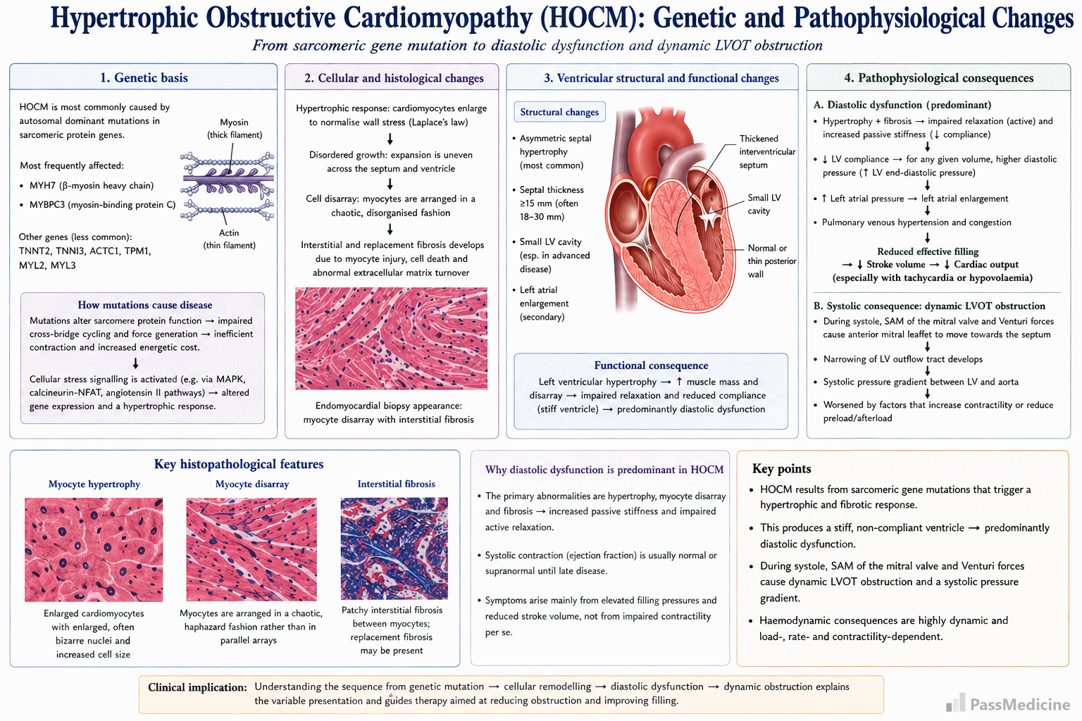
Genetics and pathophysiology in HOCM
Features
- often asymptomatic
- exertional dyspnoea
- angina
- syncope
- typically following exercise
- due to subaortic hypertrophy of the ventricular septum, resulting in functional aortic stenosis
- sudden death (most commonly due to ventricular arrhythmias), arrhythmias, heart failure
- jerky pulse, large 'a' waves, double apex beat
- systolic murmurs
- ejection systolic murmur: due to left ventricular outflow tract obstruction. Increases with Valsalva manoeuvre and decreases on squatting
- pansystolic murmur: due to systolic anterior motion of the mitral valve → mitral regurgitation
Associations
- Friedreich's ataxia
- Wolff-Parkinson White
Echo findings - mnemonic - MR SAM ASH
- mitral regurgitation (MR)
- systolic anterior motion (SAM) of the anterior mitral valve leaflet
- asymmetric hypertrophy (ASH)
ECG
- left ventricular hypertrophy
- non-specific ST segment and T-wave abnormalities, progressive T wave inversion may be seen
- deep Q waves
- atrial fibrillation may occasionally be seen
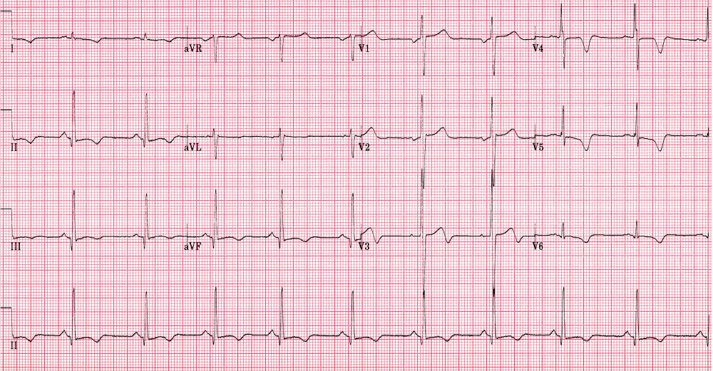
ECG of a young male with HOCM. Note the left ventricular hypertrophy and ST-T waves changes including T wave inversion - e Image used on license from Dr Smith, University of Minnesota
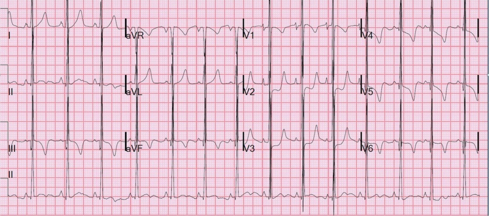
ECG showing typical changes of HOCM including LVH and T wave inversion - e Image used on license from Dr Smith, University of Minnesota
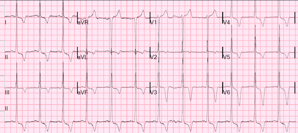
This young male patient was shown to have HOCM. Note the LVH and deep ST depression and T-wave inversions - e Image used on license from Dr Smith, University of Minnesota
+PassMedicine Notes
Hypertrophic obstructive cardiomyopathy (HOCM) is an autosomal dominant disorder of muscle tissue caused by defects in the genes encoding contractile proteins. The estimated prevalence is 1 in 500. HOCM is important as it is the most common cause of sudden cardiac death in the young.
Pathophysiology- the most common defects involve a mutation in the gene encoding #beta#-myosin heavy chain protein or myosin-binding protein C
- results in predominantly diastolic dysfunction
- left ventricle hypertrophy --> decreased compliance --> decreased cardiac output
- characterized by myofibrillar hypertrophy with chaotic and disorganized fashion myocytes ('disarray') and fibrosis on biopsy
- often asymptomatic
- exertional dyspnoea
- angina
- syncope
- typically following exercise
- due to subaortic hypertrophy of the ventricular septum, resulting in functional aortic stenosis
- sudden death (most commonly due to ventricular arrhythmias), arrhythmias, heart failure
- jerky pulse, large 'a' waves, double apex beat
- systolic murmurs
- ejection systolic murmur: due to left ventricular outflow tract obstruction. Increases with Valsalva manoeuvre and decreases on squatting
- pansystolic murmur: due to systolic anterior motion of the mitral valve --> mitral regurgitation
- Friedreich's ataxia
- Wolff-Parkinson White
- mitral regurgitation (MR)
- systolic anterior motion (SAM) of the anterior mitral valve leaflet
- asymmetric hypertrophy (ASH)
- left ventricular hypertrophy
- non-specific ST segment and T-wave abnormalities, progressive T wave inversion may be seen
- deep Q waves
- atrial fibrillation may occasionally be seen