Highlighter is on. Drag to highlight text. Click a highlight to remove it.
ALL / Haematology
Positron Emission Tomography (PET)
Positron Emission Tomography (PET)
Positron Emission Tomography (PET) is a form of nuclear imaging which uses fluorodeoxyglucose (FDG) as the radiotracer. This allows a 3D image of metabolic activity to be generated using glucose uptake as a proxy marker. The images obtained are then combined with a conventional imaging technique such as CT to decide whether lesions are metabolically active.Uses
- evaluating primary and possible metastatic disease
- assess response to treatment in certain cancers e.g. lymphoma post-chemotherapy
- assesses myocardial viability in ischaemic heart disease
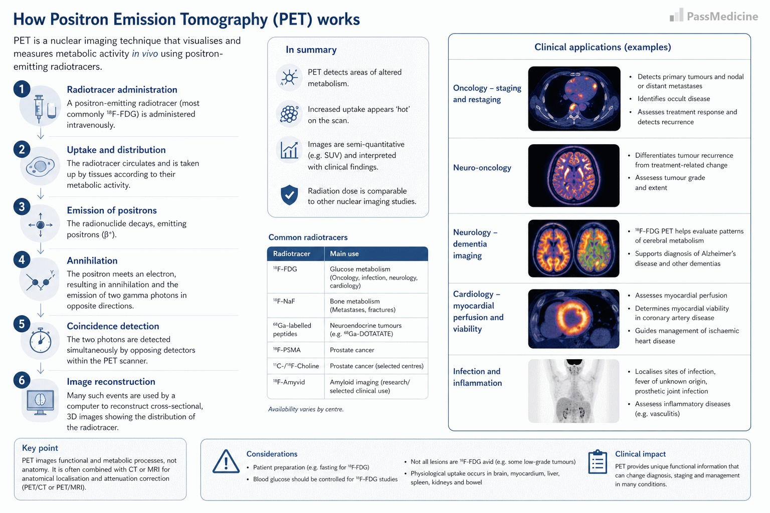
Positron Emission Tomography
+PassMedicine Notes
Positron Emission Tomography (PET) is a form of nuclear imaging which uses fluorodeoxyglucose (FDG) as the radiotracer. This allows a 3D image of metabolic activity to be generated using glucose uptake as a proxy marker. The images obtained are then combined with a conventional imaging technique such as CT to decide whether lesions are metabolically active.
Uses- evaluating primary and possible metastatic disease
- assess response to treatment in certain cancers e.g. lymphoma post-chemotherapy
- assesses myocardial viability in ischaemic heart disease