Highlighter is on. Drag to highlight text. Click a highlight to remove it.
ALL / Ophthalmology
Diabetic retinopathy
Diabetic retinopathy
Diabetic retinopathy is the most common cause of blindness in adults aged 35-65 years-old.Pathophysiology
Hyperglycaemia is thought to cause increased retinal blood flow and abnormal metabolism in the retinal vessel walls. This precipitates damage to endothelial cells and pericytes
Endothelial dysfunction leads to increased vascular permeability which causes the characteristic exudates seen on fundoscopy. Pericyte dysfunction predisposes to the formation of microaneurysms. Neovasculization is thought to be caused by the production of growth factors in response to retinal ischaemia
Classification
Patients are typically classified into those with non-proliferative diabetic retinopathy (NPDR), proliferative retinopathy (PDR) and maculopathy.
Non-proliferative diabetic retinopathy
Mild NPDR
- 1 or more microaneurysm
Moderate NPDR
- microaneurysms
- blot haemorrhages
- hard exudates
- cotton wool spots ('soft exudates' - represent areas of retinal infarction), venous beading/looping and intraretinal microvascular abnormalities (IRMA) less severe than in severe NPDR
Severe NPDR
- blot haemorrhages and microaneurysms in 4 quadrants
- venous beading in at least 2 quadrants
- IRMA in at least 1 quadrant
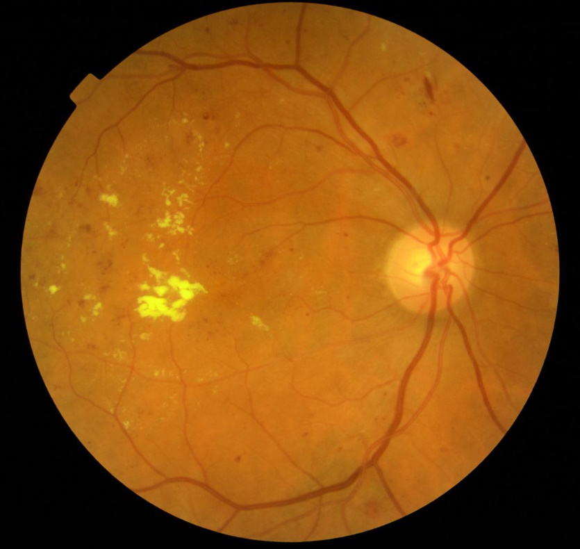
Pre-proliferative diabetic retinopathy
Proliferative diabetic retinopathy
Key features
- retinal neovascularisation - may lead to vitrous haemorrhage
- fibrous tissue forming anterior to retinal disc
- more common in Type I DM, 50% blind in 5 years
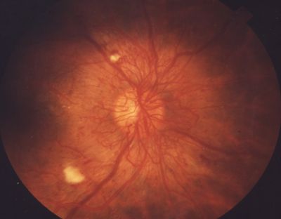
Proliferative diabetic retinopathy
Maculopathy
Key features
- based on location rather than severity, anything is potentially serious
- hard exudates and other 'background' changes on macula
- check visual acuity
- more common in Type II DM
Management
All patients
- optimise glycaemic control, blood pressure and hyperlipidemia
- regular review by ophthalmology
Maculopathy
- if there is a change in visual acuity then intravitreal vascular endothelial growth factor (VEGF) inhibitors
Non-proliferative retinopathy
- regular observation
- if severe/very severe consider panretinal laser photocoagulation
Proliferative retinopathy
- panretinal laser photocoagulation
- following treatment around 50% of patients develop a noticeable reduction in their visual fields due to the scarring of peripheral retinal tissue
- other complications include a decrease in night vision (rods are predominantly responsible for vision in low light conditions, the majority of rod cells are located in the peripheral retina), a generalised decrease in visual acuity and macular oedema
- intravitreal VEGF inhibitors
- often now used in combination with panretinal laser photocoagulation
- examples include ranibizumab
- strong evidence base suggests they both slow progression of proliferative diabetic retinopathy and improve visual acuity
- if severe or vitreous haemorrhage: vitreoretinal surgery
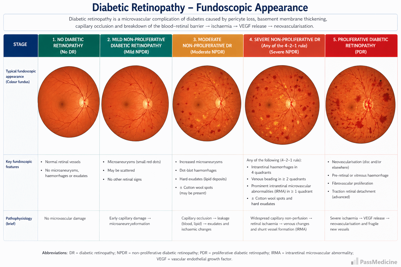
Diagram showing the different stages of diabetic retinopathy
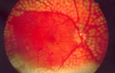
Diabetic retinopathy previously treated with panretinal laser photocoagulation
+PassMedicine Notes
Diabetic retinopathy is the most common cause of blindness in adults aged 35-65 years-old.
Pathophysiology
Hyperglycaemia is thought to cause increased retinal blood flow and abnormal metabolism in the retinal vessel walls. This precipitates damage to endothelial cells and pericytes
Endothelial dysfunction leads to increased vascular permeability which causes the characteristic exudates seen on fundoscopy. Pericyte dysfunction predisposes to the formation of microaneurysms. Neovasculization is thought to be caused by the production of growth factors in response to retinal ischaemia
Classification
Patients are typically classified into those with non-proliferative diabetic retinopathy (NPDR), proliferative retinopathy (PDR) and maculopathy.
Non-proliferative diabetic retinopathy
Mild NPDR- 1 or more microaneurysm
- microaneurysms
- blot haemorrhages
- hard exudates
- cotton wool spots ('soft exudates' - represent areas of retinal infarction), venous beading/looping and intraretinal microvascular abnormalities (IRMA) less severe than in severe NPDR
- blot haemorrhages and microaneurysms in 4 quadrants
- venous beading in at least 2 quadrants
- IRMA in at least 1 quadrant
Proliferative diabetic retinopathy
Key features- retinal neovascularisation - may lead to vitrous haemorrhage
- fibrous tissue forming anterior to retinal disc
- more common in Type I DM, 50% blind in 5 years
Maculopathy
Key features- based on location rather than severity, anything is potentially serious
- hard exudates and other 'background' changes on macula
- check visual acuity
- more common in Type II DM
Management
All patients- optimise glycaemic control, blood pressure and hyperlipidemia
- regular review by ophthalmology
- if there is a change in visual acuity then intravitreal vascular endothelial growth factor (VEGF) inhibitors
- regular observation
- if severe/very severe consider panretinal laser photocoagulation
- panretinal laser photocoagulation
- following treatment around 50% of patients develop a noticeable reduction in their visual fields due to the scarring of peripheral retinal tissue
- other complications include a decrease in night vision (rods are predominantly responsible for vision in low light conditions, the majority of rod cells are located in the peripheral retina), a generalised decrease in visual acuity and macular oedema
- intravitreal VEGF inhibitors
- often now used in combination with panretinal laser photocoagulation
- examples include ranibizumab
- strong evidence base suggests they both slow progression of proliferative diabetic retinopathy and improve visual acuity
- if severe or vitreous haemorrhage: vitreoretinal surgery