Highlighter is on. Drag to highlight text. Click a highlight to remove it.
ALL / Haematology
Thymoma
Thymoma
Thymomas are the most common tumour of the anterior mediastinum and is usually detected between the sixth and seventh decades of life.Associated with
- myasthenia gravis (30-40% of patients with thymoma)
- red cell aplasia
- dermatomyositis
- also : SLE, SIADH
Causes of death
- compression of airway
- cardiac tamponade
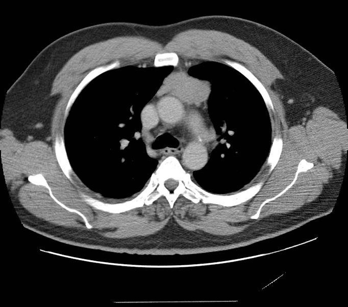
CT scan showing an anterior mediastinal mass consistent with a thymoma in a middle-aged male - e Image used on license from Radiopaedia
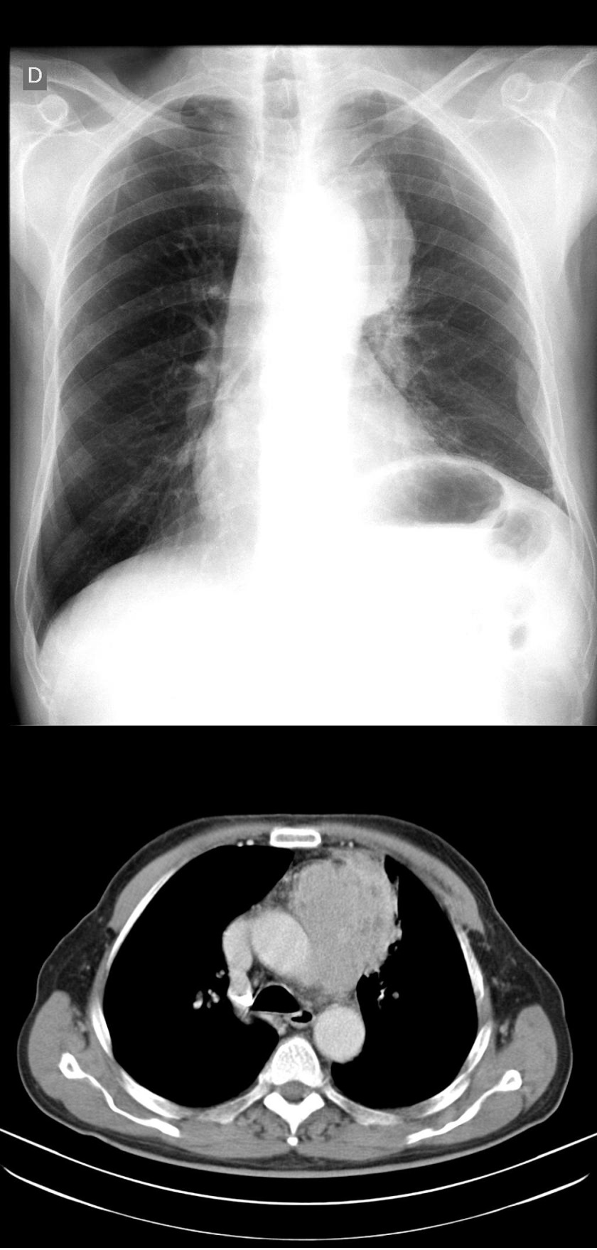
Chest x-ray and accompanying CT scan of a patient with a thymoma. In the chest x-ray there is a partially delineated mediastinal mass (anterior mediastinum) with regular borders, bulging the left upper mediastinal contour - e Image used on license from Radiopaedia
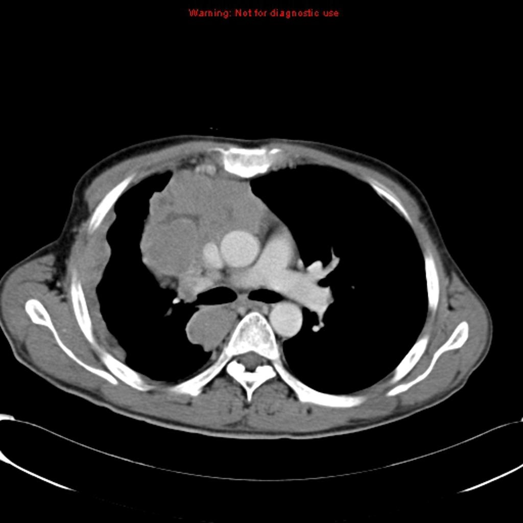
CT slice at the bifurcation of the main bronchus showing an invasive thymoma presenting as an anterior mediastinal mass - e Image used on license from Radiopaedia
+PassMedicine Notes
Thymomas are the most common tumour of the anterior mediastinum and is usually detected between the sixth and seventh decades of life.
Associated with- myasthenia gravis (30-40% of patients with thymoma)
- red cell aplasia
- dermatomyositis
- also : SLE, SIADH
- compression of airway
- cardiac tamponade