Highlighter is on. Drag to highlight text. Click a highlight to remove it.
ALL / Respiratory Medicine
Flow volume loop
Flow volume loop
A normal flow volume loop is often described as a 'triangle on top of a semi circle'- A flow–volume loop plots airflow (y-axis) against lung volume (x-axis) during forced inspiration and expiration.
- Flow volume loops are the most suitable way of differentiating between fixed and variable upper airway obstruction
- Non-invasive screening tool for suspected airway obstruction.
- Helps localise (extrathoracic vs intrathoracic) and characterise (fixed vs variable) the obstruction.
- Guides need for further imaging (CT neck/chest) or endoscopic assessment (bronchoscopy).
- Useful in pre-operative assessment (e.g. thyroid surgery) to detect subclinical tracheal narrowing.
- Can be repeated to monitor progression or response to treatment.
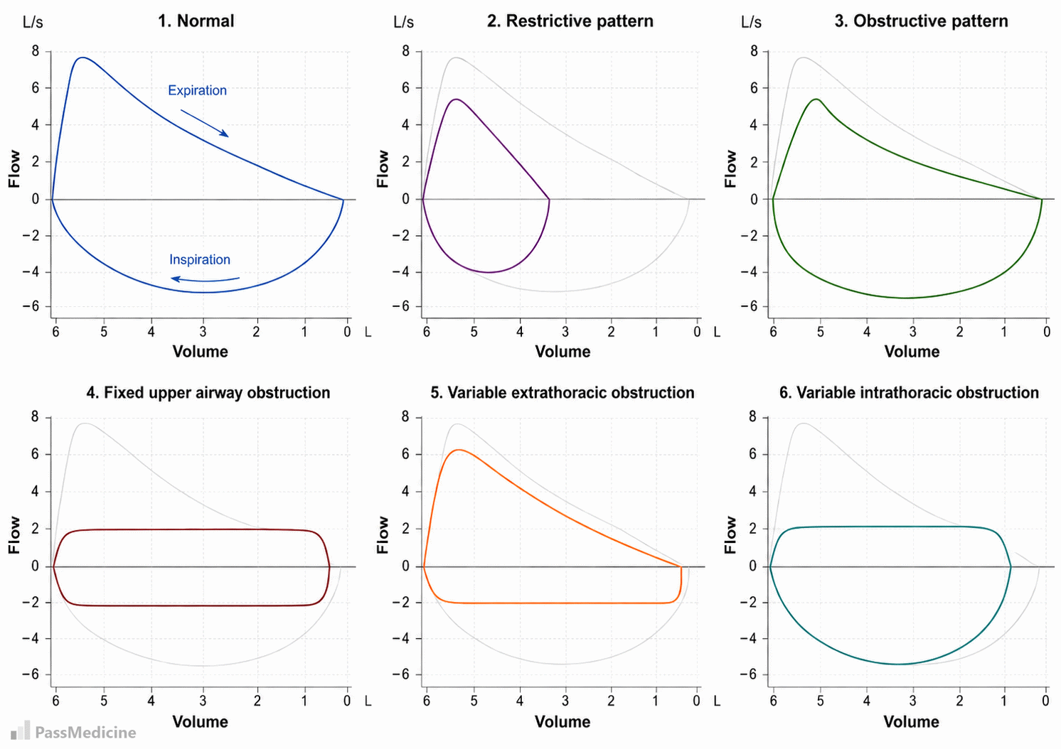
Example flow-volume loop patterns
| Fixed upper airway obstruction | Variable extrathoracic obstruction | Variable intrathoracic obstruction | |
|---|---|---|---|
| Examples | Tracheal stenosis, large goitre, tracheal tumour, fixed external compression. | Vocal cord dysfunction, laryngeal tumour, goitre pressing on trachea at neck level. | Tracheomalacia, intrathoracic goitre, proximal tracheal tumour inside the chest. |
| Appearance | Flattened inspiratory and expiratory limbs of the loop (both phases equally affected). “Box-shaped” or plateau loop. | Inspiratory limb flattened (reduced inspiratory flow). Expiratory limb usually normal. | Expiratory limb flattened (reduced expiratory flow). Inspiratory limb usually preserved. |
| Interpretation | Obstruction is constant, independent of respiratory cycle. | During inspiration, negative intrathoracic pressure narrows the extrathoracic airway, reducing flow. | During expiration, positive intrathoracic pressure narrows the intrathoracic airway, reducing flow. |
+PassMedicine Notes
A normal flow volume loop is often described as a 'triangle on top of a semi circle'
- A flow–volume loop plots airflow (y-axis) against lung volume (x-axis) during forced inspiration and expiration.
- Flow volume loops are the most suitable way of differentiating between fixed and variable upper airway obstruction
- Non-invasive screening tool for suspected airway obstruction.
- Helps localise (extrathoracic vs intrathoracic) and characterise (fixed vs variable) the obstruction.
- Guides need for further imaging (CT neck/chest) or endoscopic assessment (bronchoscopy).
- Useful in pre-operative assessment (e.g. thyroid surgery) to detect subclinical tracheal narrowing.
- Can be repeated to monitor progression or response to treatment.
“Box-shaped” or plateau loop. _ Inspiratory limb flattened (reduced inspiratory flow).
Expiratory limb usually normal. _ Expiratory limb flattened (reduced expiratory flow).
Inspiratory limb usually preserved.| Interpretation _ Obstruction is constant, independent of respiratory cycle. _ During inspiration, negative intrathoracic pressure narrows the extrathoracic airway, reducing flow. _ During expiration, positive intrathoracic pressure narrows the intrathoracic airway, reducing flow.|